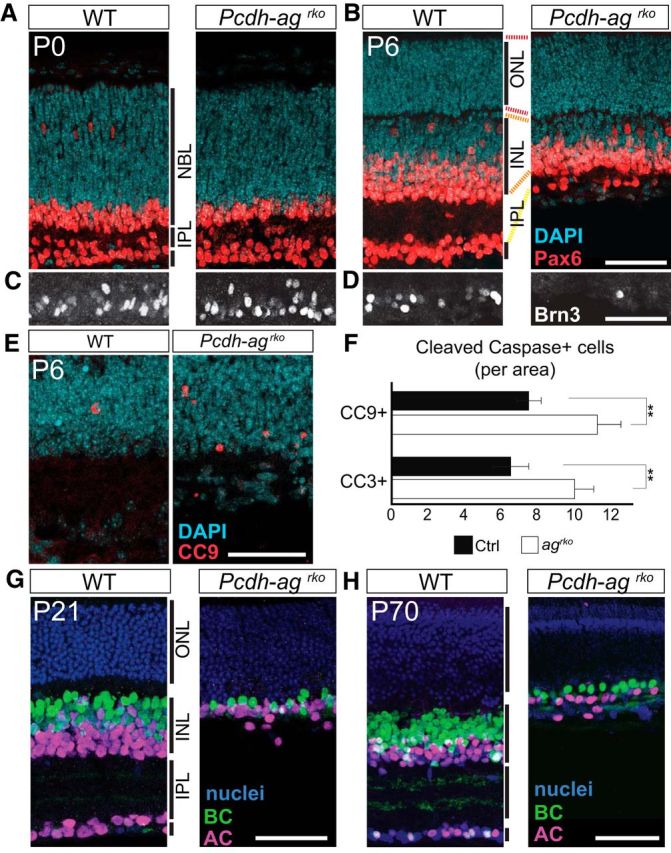
Figure 4.

Retinal cell loss from accentuated postnatal cell death in double Pcdh-ag mutant retinas. A–D, Immunolabeling of ACs (Pax6+, red; A, B) and RGCs (Brn3c+, white; C, D) reveal decline in RGC and AC populations in Pcdh-agrko retinas from P0 (A, C) to P6 (B, D) compared with WT. NBL, neuroblastic layer. E, Apoptotic cells marked by cleaved caspase-9 (CC9) in control and Pcdh-agrko retinas at P6. F, Quantifications of CC9- and cleaved caspase-3-positive cells confirmed increased apoptosis in Pcdh-agrko retinas. Data show mean ± SEM from 4–5 animals per genotype. CC3: p = 0.0051, Mann–Whitney U test, n = 18 sections. CC9: p = 0.001, Mann–Whitney U test, n = 18 sections. **p < 0.01. G, H, Comparisons of AC and BC populations in Pcdh-agrko retinas at P21 and P70 show that inner retina cell loss is not progressive with age. G, Anti-Pax6 is shown in magenta; anti-Chx10 in green, and DAPI in blue. H, Anti-AP2 is shown in magenta; anti-Chx10 in green, and TO-PRO in blue. Dashed lines indicate retinal layers. Scale bars, 50 μm.
