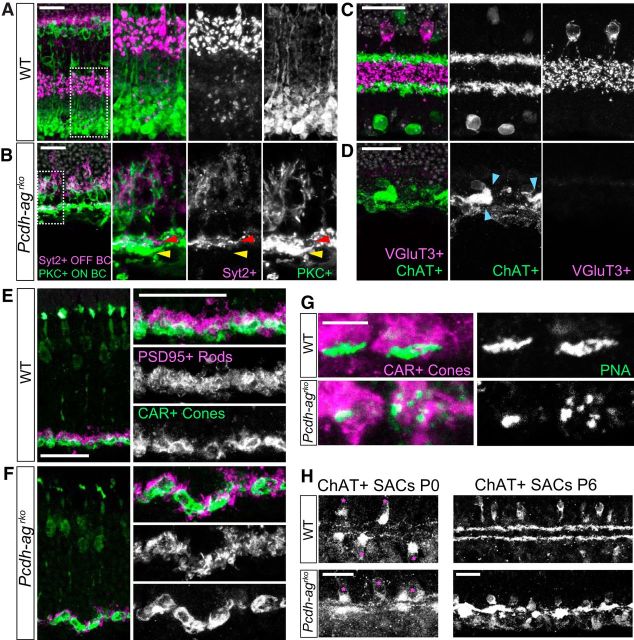
Figure 5.
Laminar organization of the IPL and OPL is not maintained in the absence of Pcdha and Pcdhg clusters. A, B, Sections of inner retina showing laminar segregation of OFF cone bipolar (Syt2+, magenta) and ON rod bipolar axon terminals (PKCα+, green) in IPL of P21 WT (A) and Pcdh-agrko retinas (B). White boxes in left panels indicate regions shown at higher magnification in the other panels. Arrows indicate OFF (red) and ON (yellow) BC terminals, which are intermingled in Pcdh-agrko. C, D, Laminar segregation of AC processes: vGluT3+ narrow field ACs (magenta) form elaborate processes between two ChAT+ SAC layers (green) in WT (C) at P21. In Pcdh-agrko retinas (D), vGluT3+ ACs are absent and ChAT+ layers are collapsed. Blue arrows denote clumping of SAC dendrites. E, F, Sections of outer retina showing laminar organization of rod (PSD95+, magenta) and cone (CAR+, green) terminals in OPL. E, WT. F, Rod spherules are displaced relative to cone pedicles in Pcdh-agrko. G, Lectin PNA labeling show fragmentation of cone synaptic structures in Pcdh-agrko retinas. H, SAC processes are stratified in WT and Pcdh-agrko retina at P0 (left), but are collapsed in Pcdh-agrko retina at P6 (right). Scale bars: A–F, H, 25 μm; G, 5 μm.

