Figure 7.

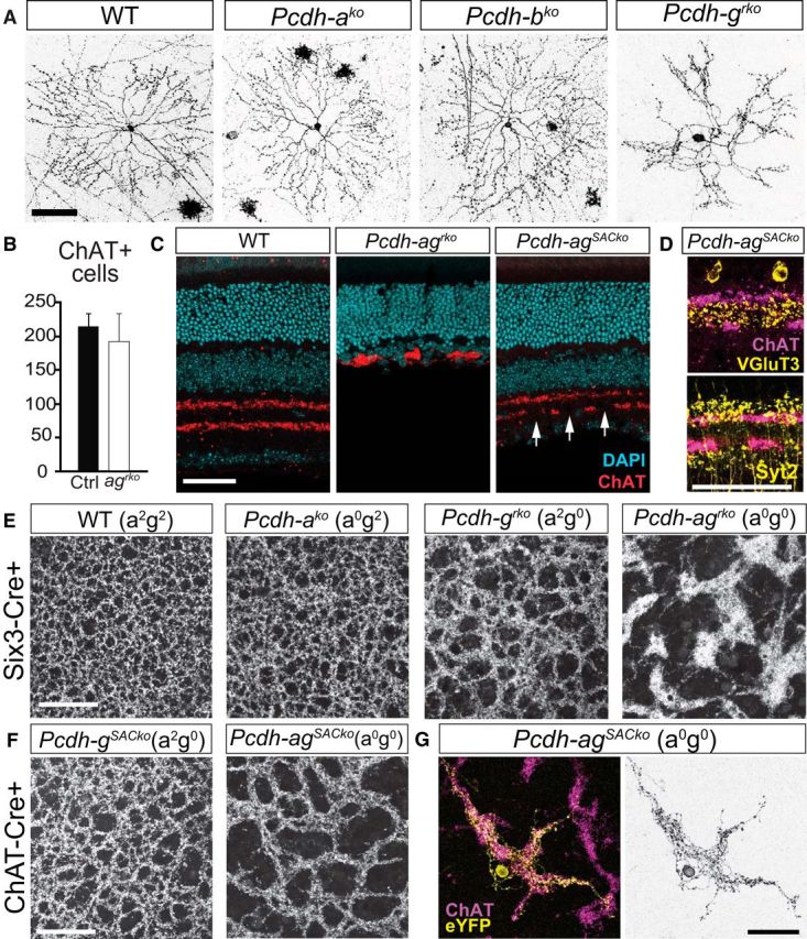
The Pcdha and Pcdhg clusters cooperate to promote dendrite self-avoidance. A, Single SACs labeled with AAV-membrane-RFP in WT, Pcdhako, Pcdhbko, and Pcdhgrko retinal whole mounts. B, Numbers of ChAT+ SACs in WT and Pcdh-agrko retinas. Bars show means ± SEM. n = 17 areas were analyzed, 3 animals per genotype, Student's t test, t(32) = 2.0, p = 0.059. C, SAC dendritic layers in WT, Pcdh-agrko, and Pcdh-agSACko [Pcdh-aconexldel; gf x Chat-cre] retinas labeled with DAPI (cyan) and anti-ChAT (red). Arrows indicate gaps in the SAC layers despite preservation of the inner retina in Pcdh-agSACko retina. D, Laminar segregation of vGluT3+ AC processes (yellow, top) and Syt2+ BC terminals (yellow, bottom) is intact in Pcdh-agSACko retina (ChAT+ SAC layers, magenta). E, F, Whole-mount retina preparation showing ChAT-labeled plexus formed by SAC dendrites in WT, single Pcdh-ako and Pcdh-grko mutants, and double Pcdh-agrko mutants (E) and SAC-specific Pcdh-gSACko and Pcdh-agSACko mutants (F). Note larger gaps and increased fasciculation of SAC plexus in double mutant Pcdh-ag compared with WT or single Pcdhg mutant retinas. G, Morphology of a single SAC from Pcdh-agSACko retina labeled with eYFP (yellow) and SAC plexus labeled with anti-ChAT (magenta). Scale bars, 50 μm.
