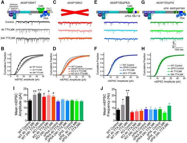
Figure 2.
Rapid homeostatic synaptic potentiation in hippocampal neurons requires AKAP150-anchoring of both PKA and CaN: disruption by knock-out of AKAP150 (KO) and genetic deletion of PKA-anchoring (ΔPKA) or CaN-anchoring (ΔPIX). A, Representative mEPSC recordings and (B) cumulative plots of mEPSC amplitudes showing scaling-up induced by 4 or 24 h TTX (2 μm) with MK801 (10 μm) present for the last 3 h (4 h TTX/MK or 24 h TTX/MK) in 15–16 DIV WT mouse hippocampal neurons (data are reproduced from Fig. 1A and B). C, Representative mEPSC recordings and (D) cumulative plots of mEPSC amplitudes showing that scaling-up induced by 4 or 24 h TTX/MK treatment is absent in neurons from AKAP150 KO mice. Under basal conditions AKAP150 KO neurons already exhibit a rightward shift in mEPSC amplitude distribution toward larger amplitudes compared with WT controls (black line) and the 4 and 24 h TTX/MK treatments fail to produce any additional shifts. E, Representative mEPSC recordings and (F) cumulative plots of mEPSC amplitudes showing that scaling-up induced by 4 or 24 h TTX/MK is absent in neurons from PKA anchoring-deficient AKAP150ΔPKA mice. G, Representative mEPSC recordings and (H) cumulative plots of mEPSC amplitudes showing that scaling-up induced by 4 or 24 h TTX/MK is absent in neurons from CaN anchoring-deficient AKAP150ΔPIX mice. Bar graph summaries of data in A–H showing the 4 and 24 h TTX/MK conditions induce significant increases in mean mEPSC (I) amplitude and (J) frequency in hippocampal neurons from WT but not from AKAP150 KO, ΔPKA, or ΔPIX mice. *p < 0.05, **p < 0.01 to corresponding Controls by one-way ANOVA; #p < 0.05 to WT Control by one-way ANOVA. Error bars indicate SEM.

