
Figure 5.
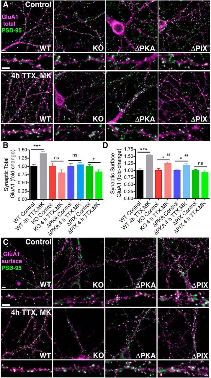
Induction of rapid homeostatic synaptic potentiation increases synaptic GluA1 localization in hippocampal neurons cultured from WT but not AKAP150 KO, ΔPKA, or ΔPIX mice. A, Representative images showing total GluA1 (magenta) and PSD-95 (green) colocalization (white) in 16 DIV hippocampal neurons cultured from WT, AKAP150 KO, ΔPKA, or ΔPIX mice under control conditions or after induction of rapid homeostatic scaling-up with 4 h TTX/MK treatment. Bottom, Magnifications of dendrites. Scale bars, 5 μm. B, Quantification of fold-change in total synaptic GluA1 colocalization with PSD-95 from images as in A using Pearson's correlation (r) shows that induction of homeostatic scaling-up with 4 h TTX/MK significantly increases GluA1/PSD-95 colocalization in WT (***p < 0.001 to Control by unpaired t test) but not KO or ΔPKA mouse neurons (both NS p > 0.05 to Controls by unpaired t test). Total GluA1/PSD-95 colocalization actually decreases slightly in ΔPIX mouse neurons after 4 h TTX/MK (*p < 0.05 to Control by unpaired t test). Error bars indicate SEM. C, Representative images showing surface GluA1 (magenta) and PSD-95 (green) colocalization (white) in 16 DIV hippocampal neurons cultured from WT, AKAP150 KO, ΔPKA, or ΔPIX mice under control conditions or after induction of rapid homeostatic scaling-up with 4 h TTX/MK treatment. Bottom, Magnifications of dendrites. Scale bars, 5 μm. D, Quantification of fold-change in synaptic surface GluA1 colocalization with PSD-95 from images as in C using Pearson's correlation (r) shows that induction of homeostatic scaling-up with 4 h TTX/MK treatment increases surface GluA1/PSD-95 colocalization in WT mouse neuron (***p < 0.0001 to Control by unpaired t test) significantly more than in KO or ΔPKA mouse neurons (both *p < 0.05 to corresponding Controls by unpaired t test and ##p < 0.01 to WT 4 h TTX/MK by one-way ANOVA). Surface GluA1/PSD-95 colocalization is unchanged in ΔPIX mouse neurons after 4 h TTX/MK (NS p > 0.05 to Control by unpaired t test). Error bars indicate SEM.