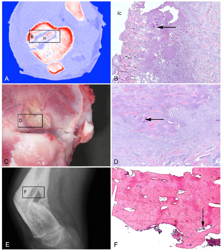
Figure 1.
Bone tissue used in the present study. A: Porcine model of implant-associated osteomyelitis. CT scan after five days of inoculation demonstrating the tibial implant cavity (ic) used for injection of S. aureus and insertion of a small steel implant. B: Histology of picture A demonstrating bacteria (arrow) in the peri-implant tissue adjacent to the implant cavity (ic). HE. C: Porcine model of haematogenous osteomyelitis euthanized fifteen days after inoculation. A lesion is shown in the right femoral metaphysis. D: Histology of picture C demonstrating bacteria (arrow) centrally in the lesion. HE. E: X-ray of a child with haematogenous osteomyelitis and pathological fracture in the right femur. F: Histology of picture E demonstrating bacteria (arrow) in a cortical sequester. HE.

