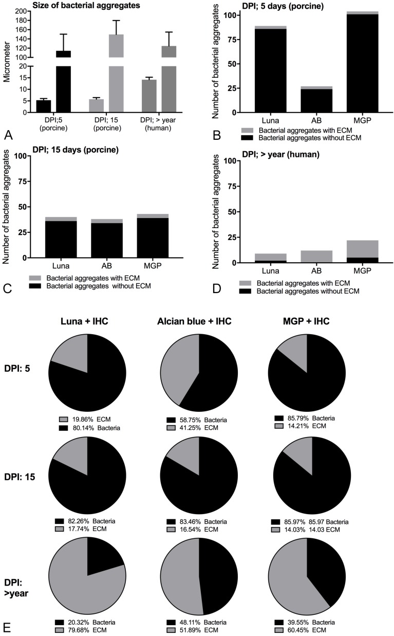
Figure 4.
Composition and size of biofilm forming infections in porcine and human bone tissue. A: Size of the three smallest and three largest bacterial aggregates seen with immunohistochemistry towards S. aureus, Mean ± SD. B-D: Number of bacterial aggregates with and without a visible extracellular biofilm matrix at 100x magnification following combined histochemistry with immunohistochemistry. The x-axis shows the histochemical stains. E: Percentage of bacterial cells and extracellular matrix in single representative S. aureus biofilm aggregates. AB: Alcian Blue pH3, MGP: Methyl-pyronin green, DPI: days post infection, ECM: extracellular matrix.

