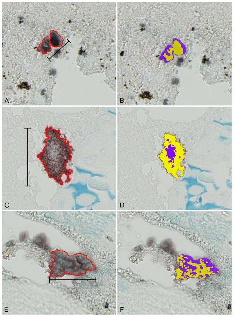
Figure 5.
Percentage of bacteria and extracellular matrix in S. aureus biofilm infections. Left column: combined immunohistochemistry (towards S. aureus) and Alcian blue pH3 staining of representative bacterial biofilms (red outline) displaying both bacteria (red/brown) and extracellular matrix(blue). Right column demonstrates calculations of % bacterial cells (yellow) and extracellular matrix (purple). A+B: Bone tissue from a porcine model of S. aureus implant-associated osteomyelitis infected for five days. Bar = 40 μm. C+D: Bone tissue from a porcine model of S. aureus haematogenous osteomyelitis infected for fifteen days. Bar = 78 μm. E+F: Bone tissue from a patient suffering from S. aureus haematogenous osteomyelitis for more than one year. Bar = 72 μm.

