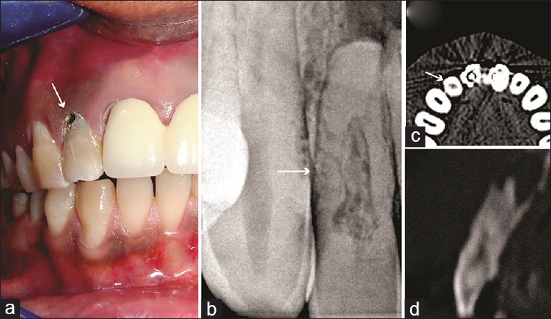
Figure 1.

(a) A preoperative photograph of tooth 12 showing discolored tooth 12 with a defect at cervical third region (arrow). (b) A preoperative radiograph suggestive of obliterated coronal pulp chamber and enlarged space and concomitant calcific depositions with middle-third of root canal (arrow). (c) The axial section at the level of middle-third of root reveals internal root resorption (arrow). (d) The sagittal section revealed a resorptive defect with concomitant radiopaque deposition
