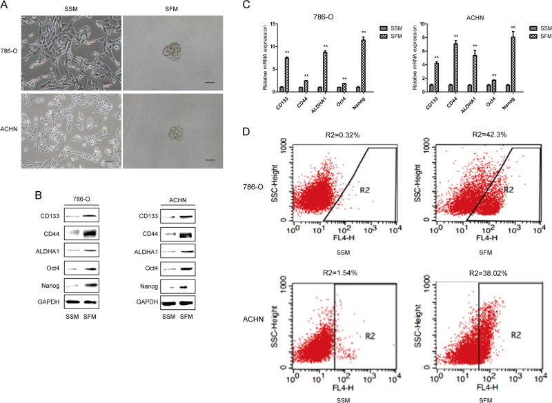
Fig. 1. Tumorsphere formation assay of renal CSCs in SFM condition.
786-O and ACHN cells were grown in serum-supplied medium (SSM) and serum-free medium (SFM) for 5 days, respectively. a Representative images of tumorspheres. Bar, 100 μm. b The protein expression of renal CSCs markers (CD133, CD44, ALDHA1, Oct4, and Nanog) was measured by Western blotting. c The mRNA expression level of renal CSCs was measured by qRT-PCR. d The number of CD133+ cells in 786-O and ACHN cells was detected by flow cytometry. Data are expressed as mean ± standard deviation (SD). *p < 0.05, **p < 0.01 compared with SSM group

