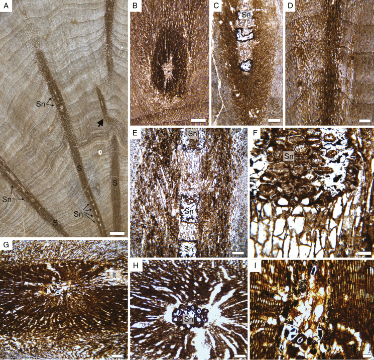
Fig. 6.
Triassic trees with epicormic shoots from Gordon Valley: shoot anatomy. (A) View of a cross-section of wood showing several epicormic traces (S) with an oblique to horizontal trajectory. The shoot on the right is branching (arrow). Note regularly spaced sclerotic nests in the pith of the shoots (Sn). 18,292 Dbot1 (slide 30,292). (B) Cross-section of a shoot at a level with no sclerotic nest in the pith. 18,294 Btop1. (C) Oblique view of a shoot showing some sclerotic nests in the pith. 18,292 Cbot1 (slide 30,291). (D) Oblique view of a shoot for which only the secondary xylem is visible. Note that there are no conspicuous frost rings associated with the trace. 18,291 Cbot1 (slide 30,291). (E) Detail of a shoot seen in longitudinal section, with an alternation of parenchyma and sclerotic nests in the pith. 18,292 Cbot1 (slide 30,291). (F) Detail of the pith on (E) 18,292 Cbot1 (slide 30,291). (G) Shoot producing a trace to a lateral organ (lt). 18,294 A6 (slide 30,286). (H) Transverse-oblique section of a shoot showing a large sclerotic nest (Sn) in the pith. 18,294 B3 (slide 30,289). (I) Longitudinal section of a shoot showing the pith and secondary xylem with uniseriate pits on the tracheid walls. 18,294 A6 (slide 30,286). Scale bars: (A) 1 cm; (B–D) 500 µm; (E, G) 250 µm; (H) 100 µm; (F) 50 µm.

