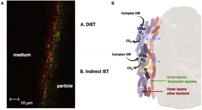
FIGURE 7.
(A) FISH image from the section of the biofilm developed on an activated carbon particle, using Eubacteria probe (red signal), and Geobacter cluster probe (green signal) (probes combination 3). The left side of the red signal corresponds to the outermost environment (ME-FBR medium), whereas the right side of the green signal corresponds to the surface of a fluidized activated carbon particle. This image is representative of one out of 30 sections taken from the biofilm developed on a surface of ca. 50 × 100 μm. (B) Proposed microbial electron transfer mechanisms among the different microbial communities colonizing the particles and toward the fluidized anode.

