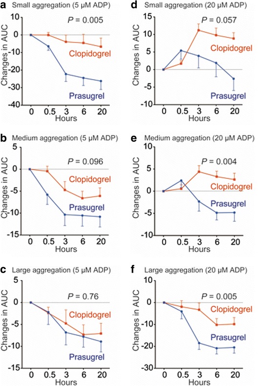
Fig. 4.

Platelet aggregation as assessed by the laser light scattering method after the loading dose (LD). Platelets in platelet-rich plasma obtained at the indicated time points after the LD were stimulated with 5 μM adenosine diphosphate (ADP) (a–c) or 20 μM ADP (d–f). Small (a, d), medium (b, e), and large (c, f) platelet aggregate formation was measured by the laser light scattering method. This formation is expressed as changes in the area under curve (AUC) of each platelet aggregate number during 5 min ( x 106 V/min). Blue lines and red lines represent the LD of prasugrel (20 mg) and that of clopidogrel (300 mg), respectively. Values are means ± SEM. Differences in the time course between groups were determined by repeated measures ANOVA
