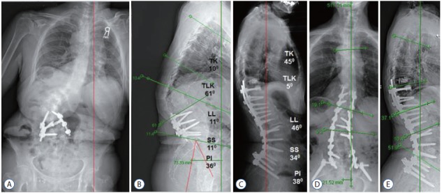
Fig. 2.

A 72-year-old female with back pain and poor posture. Scoliosis AP radiographs (A) and lateral radiographs (B) reveal with poor balance and native PI of 36°. Target lumbar lordosis was 54°±7° (47° to 61°). Mismatch between target LL and current LL was 34°±7° (27° to 41°). Operative plan was a single PSO to obtain 35° versus six posterior column osteotomies (PCOs) to obtain 30°–42°. The decision was made to proceed with PSSIF T10–S1–Pelvis with PLIF at L5–S1 and PCOs at T10–11, T11–12, T12–L1, L1–2, L2–3, and L5–S1. Scoliosis lateral at 8 weeks (C) shows TK of 45° and LL of 46°. Perfect sagittal balance was achieved. Scoliosis AP (D) and lateral (E) in 2 years. Perfect sagittal balance was maintained. TK : thoracic kyphosis, TLK : thoracolumbar kyphosis, LL : lumbar lordosis, SS : sacral slope, PI : pelvic incidence, AP : anterior posterior, PSO : pedicle subtraction osteotomy, PSSIF : posterior spinal segmental instrumentation and fusion, PLIF : posterior lumbar interbody fusion.
