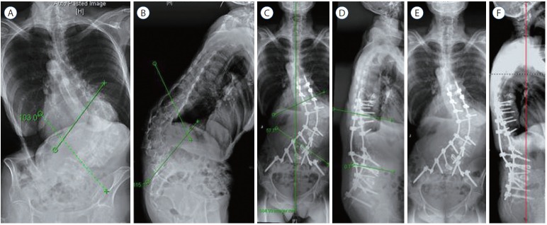
Fig. 4.
A 73-year-old female with back pain and poor posture. Scoliosis AP radiographs (A) and lateral radiographs (B) reveal with massive fixed kyphoscoliosis of 103° scoliosis and 115° kyphosis. Operative plan was PSSIF T9–S1–Pelvis with L1 PVCR and PLIF at L4–5 and L5–S1 with multilevel PCOs. Scoliosis AP (C) and lateral (D) at 8 weeks show correction of scoliosis to 47° and correction of kyphosis to 114°. Scoliosis AP (E) and lateral (F) at 4 years. Perfect sagittal balance was maintained. AP : anterior posterior, PSSIF : posterior spinal segmental instrumentation and fusion, PVCR : posterior vertebral column resections, PLIF : posterior lumbar interbody fusion, PCOs : posterior column osteotomies.

