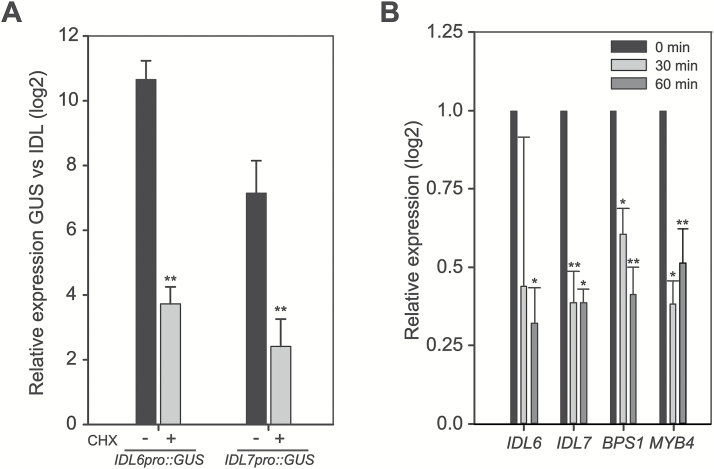
Fig. 2.
Rapid turnover of IDL6 and IDL7 mRNA. (A) Ten-day-old reporter lines (n=3) expressing GUS under control of the IDL6 and IDL7 promoters were analysed for GUS and IDL6/IDL7 expression through qRT–PCR with (two independent lines) or without (four independent lines) CHX treatment (10 µg ml–1, 3 h). The mean and SD of the expression ratio (log2) between GUS and IDL6/IDL7 for each construct is shown. (B) mRNA decay analysis showing the relative gene expression level of IDL6 and IDL7 at 30 min and 60 min after adding of cordycepin (1 mM) to the media. BPS1 and MYB4 were included as controls (n=4). Statistical differences (REST analysis: *P-value <0.05; **P-value <0.01) between the time point samples and control are indicated. Error bars indicate SDs.

