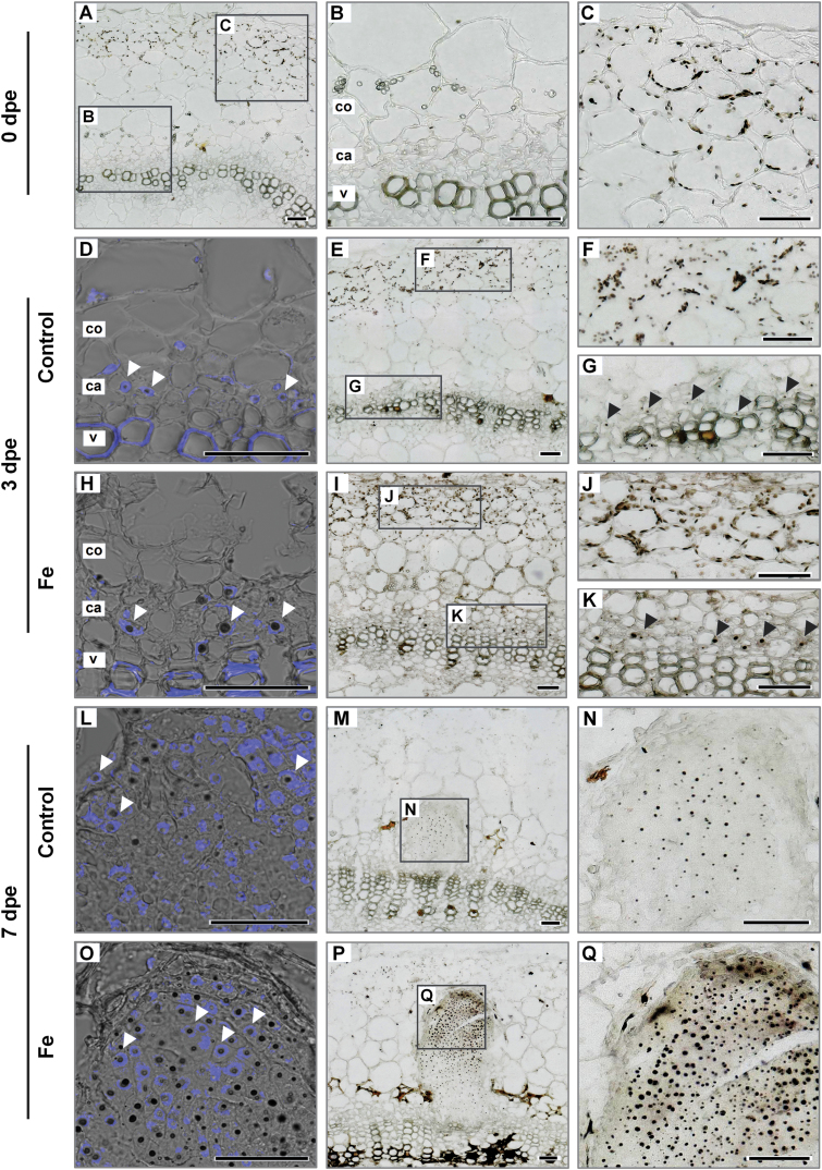
Fig. 10.
Localization of Fe by Perls’ Prussian blue–DAB method in the stem base of Petunia hybrida cuttings in response to Fe supply during adventitious root formation. Cross sections from 1–4 mm of the stem base are shown 0 dpe (A–C), 3 dpe (D–K) and 7 dpe (L–Q) in cuttings grown without nutrients (A–G; L–N) or with Fe (H–K; O–Q). Black arrowheads show Fe localization in cambial cells 3 dpe. Additional staining with DAPI (D, H, L, O) indicates nuclear localized Fe in meristematic cells (white arrowheads). ca. cambium; co. cortex; pp. pith parenchyma; v. vessels of xylem. Scale bar, 50 µm.

