Fig. 2.
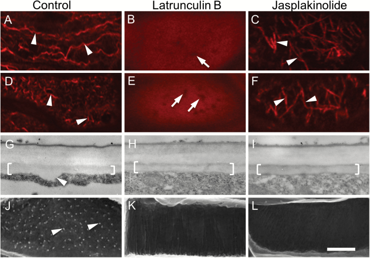
Effect of actin depolymerizing and stabilizing drugs on the actin network, and deposition of the uniform wall layer and WI papillae, in trans-differentiating adaxial epidermal cells of cultured V. faba cotyledons. Cotyledons were cultured for 4 h in the absence (A, D, G, J) or presence of 100 nM of the actin-depolymerizing drug latrunculin B (B, E, H, K) or 100 nM of the actin-stabilizing drug jasplakinolide (C, F, I, L) together with 100 μM, aminoethoxyvinylglycine (AVG) to inhibit initiation of trans-differentiation to a TC morphology (A–C). Thereafter, cotyledons were transferred to AVG-free media and cultured for a further 15 h (D–L). (A–F) Representative CLSM images of the actin network visualized with Rhodamine-phalloidin at the outer periclinal cell wall/cytoplasmic interface (Z-depth 0 nm). Comparing (A) with (B) and (C) illustrates the depolymerizing (B) and stabilizing (C) effects of latrunculin B and jasplakinolide on the actin network after the 4-h pre-treatment. By 15 h of culture, following induction of TC trans-differentiation, these effects remain unchanged (E, F) except for actin bundles being re-orientated to a more transverse configuration in the jasplakinolide treatment (arrowheads in F). In the absence of the drugs (D), the actin network was remodelled by fragmentation into short actin bundles (arrowheads in D, compare those in A). Actin-free zones (arrows in B, E) are likely to be occupied by mitochondria (1.17 ± 0.03 µm in length; diameter 0.46 ± 0.01 µm; n=100). (G–I) Representative TEM images of transverse sections of the outer periclinal wall of adaxial epidermal cells illustrating the uniform wall layer (brackets). The arrowhead in (G) indicates a WI papilla. (J–L) Representative SEM images of the cytoplasmic face of the outer periclinal wall of adaxial epidermal cells. Note WI papillae in (J) (arrowheads) but no papillae in (K) and (L). Scale bar represents 5 μm in (A–F) and 500 nm in (G–I).

