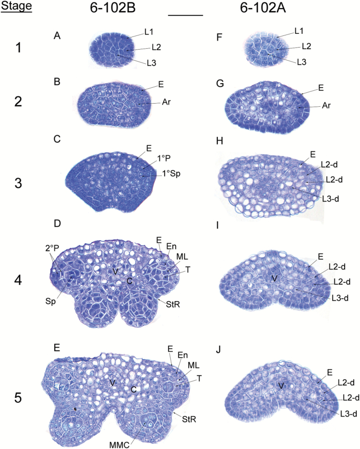
Fig. 2.
Anther development defects in the hau CMS line in B. juncea. The images are semi-thin sections from (A–E) the hau CMS maintainer line (6-102B) and (F–J) the hau CMS line (6-102A) showing anther development from stages 1–5. Abbreviations: Ar, archesporial cell; E, epidermis; En, endothecium; L1, L2, and L3, the three cell layers of the stamen primordia; L2-d and L3-d, the L2- and L3-derived cells; ML, middle layer; MMC, microspore mother cell; Sp, sporogenous cells; StR, stomium region; T, tapetum; V, vascular tissue; 1°P, primary parietal layer; 1°Sp, primary sporogenous layer; and 2°P, secondary parietal cell layers. Scale bar =25 µm. (This figure is available in colour at JXB online.)

