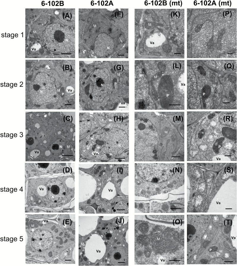
Fig. 3.
TEM analysis of anthers from the hau CMS maintainer line (6-102B) and the hau CMS line (6-102A) at different stages. Anther development from stages 1–5 in 6-102B (A–E) and in 6-102A (F–J). The mitochondrial structures of the cells from (A–E) and (F–J) are shown in (K–O) and (P–T), respectively. Abbreviations: M, mitochondria; P, plastid; N, nucleus; and Va, vacuole. Scale bars: (D) 0.2 µm, (B, F, H, L, P, R) 0.5 µm, (C, E, G, J, N, Q, T) 1 µm, (A, K, M, O, S) 2 µm, and (I) 5 µm.

