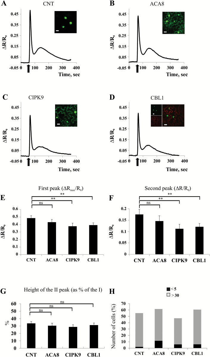
Fig. 4.
Ca2+ signatures induced by leaf mechanical wounding in tobacco leaves expressing each of the tested proteins alone. (A–D) Nuclear Ca2+ concentration monitoring in N. benthamiana leaf cells transiently expressing NUP::YC3.6 alone (CNT, A) or co-expressing NUP::YC3.6 and ACA8::GFP (B), NUP::YC3.6 and CIPK9::GFP (C), or NUP::YC3.6 and CBL1::OFP (D). Leaves were challenged with wounding (arrow), and FRET variations (normalized FRET cpVenus/CFP ratio reported as ΔR/R0) in single cells surrounding the wounded site were observed for ~400 s at 2 s intervals. Traces are the averages from the analysis of at least 25 independent cells. Insets: single plane confocal images of N. benthamiana epidermal cells from leaves used for wounding experiments, showing the simultaneous expression of the different expressed fluorescent proteins. (E and F) Comparison and statistical analysis of the height of the first and second peaks of the Ca2+ transients (as determined by single-cell analysis) reported as the normalized ΔR/R0 (±SEM), measured in the different tested conditions. (G) Mean height of the second peak expressed as a percentage of the height of the first peak (±SEM). Asterisks indicate statistically significant differences (**P<0.05, ns=not significant) calculated using Student’s t-test. (H) Comparison of the number of epidermal cell nuclei in which the height of the second peak is <5% or >30% of the height of the first one in N. benthamiana leaves infiltrated with the different combinations of A. tumefaciens harbouring the different plasmids.

