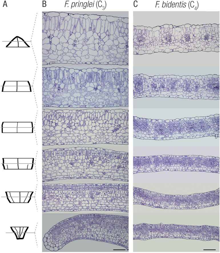
Fig. 2.
Within-leaf sampling indicates the gradual maturation gradient in leaf anatomy for C3 and C4Flaveria speciecs. (A) Representative leaf outline illustrating leaf sampling. Representative transverse sections from C3Flaveria pringlei (B) and C4Flaveria bidentis (C) from base (bottom) to tip (top). Note the gradual expansion of cells, increased vacuolization, and clearer delineation of both mesophyll and bundle sheath cells from base to tip. Scale bars represent 100 μm.

