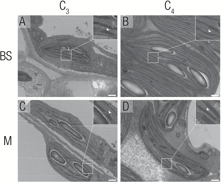
Fig. 8.
Chloroplast ultrastructure of mesophyll and bundle sheath cells from C3F. pringlei and C4F. bidentis. Transmission electron microscopy was used to investigate chloroplast ultrastructure. (A, B) Bundle sheath chloroplasts from C3Flaveria pringlei and C4Flaveria bidentis. (C, D) Mesophyll chloroplasts from C3F. pringlei and C4F. bidentis. Little granal stacking was seen in bundle sheath chloroplasts from F. bidentis (B) whereas it could be observed in C3 bundle sheath and in all mesophyll chloroplasts. Insets show a close-up of thylakoids, with white arrowheads indicating thylakoid stacks (A, C, D) or the lack of extensive stacking (B). Scale bars represent 500 nm.

