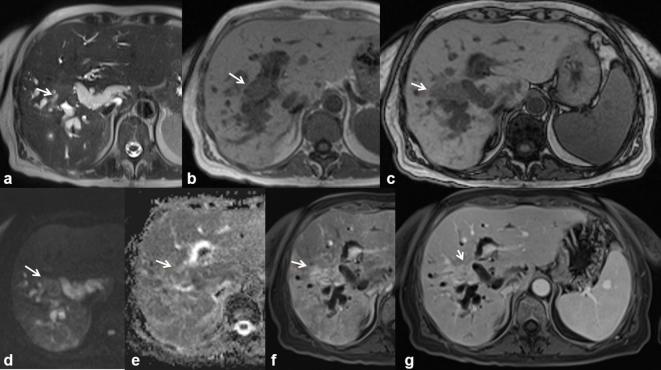
Figure 12.
56-year-old female. Pancreatic cancer with peribiliary liver metastases. The metastatic tissue is hyperintense (arrow) on T2-W HASTE images (a) and hypointense on T1 W images (b, in-of-phase T1 W and c out-of-phase T1 W). Diffusivity is restricted on DW images (d, b800;e, ADC map) with a mean ADC value of 1.28 × 10⁻³ mm2 s–1. Vascularized tissue (arrow) (f, arterial and g, portal-phase T1 W VIBE images).

