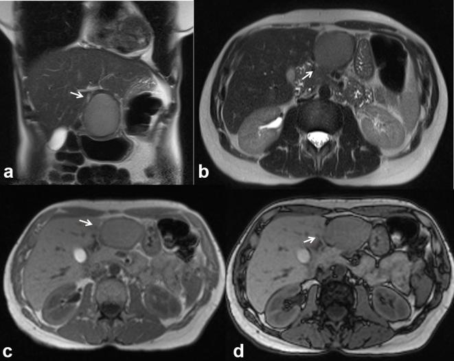
Figure 4.
The same patient of Figure 3. MRI demonstrates a complex lesion with proteinaceous content. The lesion is iso-hyperintense (arrow) on T2 W (HASTE, (a, coronal view; b, axial view) and on T1 W (c, in-of-phase and d, out-of-phase axial image). HASTE, half-Fourier acquisition single-shot turbo spin-echo.

