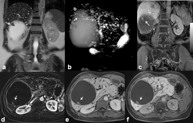
Figure 6.
50-year-old female. Cystadenocarcinoma. Cystic mass with a solid, enhancing mural nodule (arrow). The mural nodule appears hyperintense (arrow) on T2 W images (a, HASTE T2 W coronal view; b, single-shot cholangiographic image), hyperintense (arrow) on hepatospecific phase on contrast study (c, VIBE T1 W FS image coronal plane), hypeintense on contrast-enhanced arterial-phase axial image (d, VIBE T1 W FS axial plane), contrast-enhanced portal-phase axial image (e) and contrast-enhanced hepatobiliary phase axial image (f). HASTE, half-Fourier acquisition single-shot turbo spin-echo.

