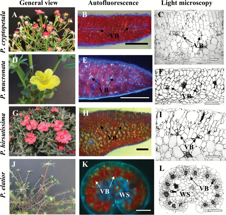
Fig. 1.
General view of Portulaca species (left panels, A, D, G, J), distribution of chlorenchyma under the fluorescent microscope (middle panels, B, E, H, K), and light microscopy of leaf cross sections (right panels, C, F, I, L). P. cryptopetala (B, C), P. mucronata (E, F) and P. hirsutissima (H, I), all have C3 like dorsoventral type leaf anatomy. In P. cryptopetala and P. mucronata (B, C, E, F), all vascular bundles (VB), including the main vein, are distributed in the median paradermal plane; red fluorescence from chlorophyll is distributed more or less evenly through the leaf mesophyll (B, E). In P. hirsutissima (H, I), VB are positioned close to the adaxial side while the main vein is in the center of the leaf below lateral veins; red chlorophyll fluorescence is concentrated in the adaxial palisade mesophyll layers with water storage (WS) spongy mesophyll cells in the middle of the leaf (H). P. elatior has Pilosoid type of anatomy (K, L) with peripheral distribution of VB with each vein surrounded by two Kranz chlorenchyma layers; WS tissue is around the main vein. VB, vascular bundles; WS, water storage tissue. Scale bars: 1 mm for B; 200 µm for C, F, I, L; 0.5 mm for E, H, K.

