Fig. 2.
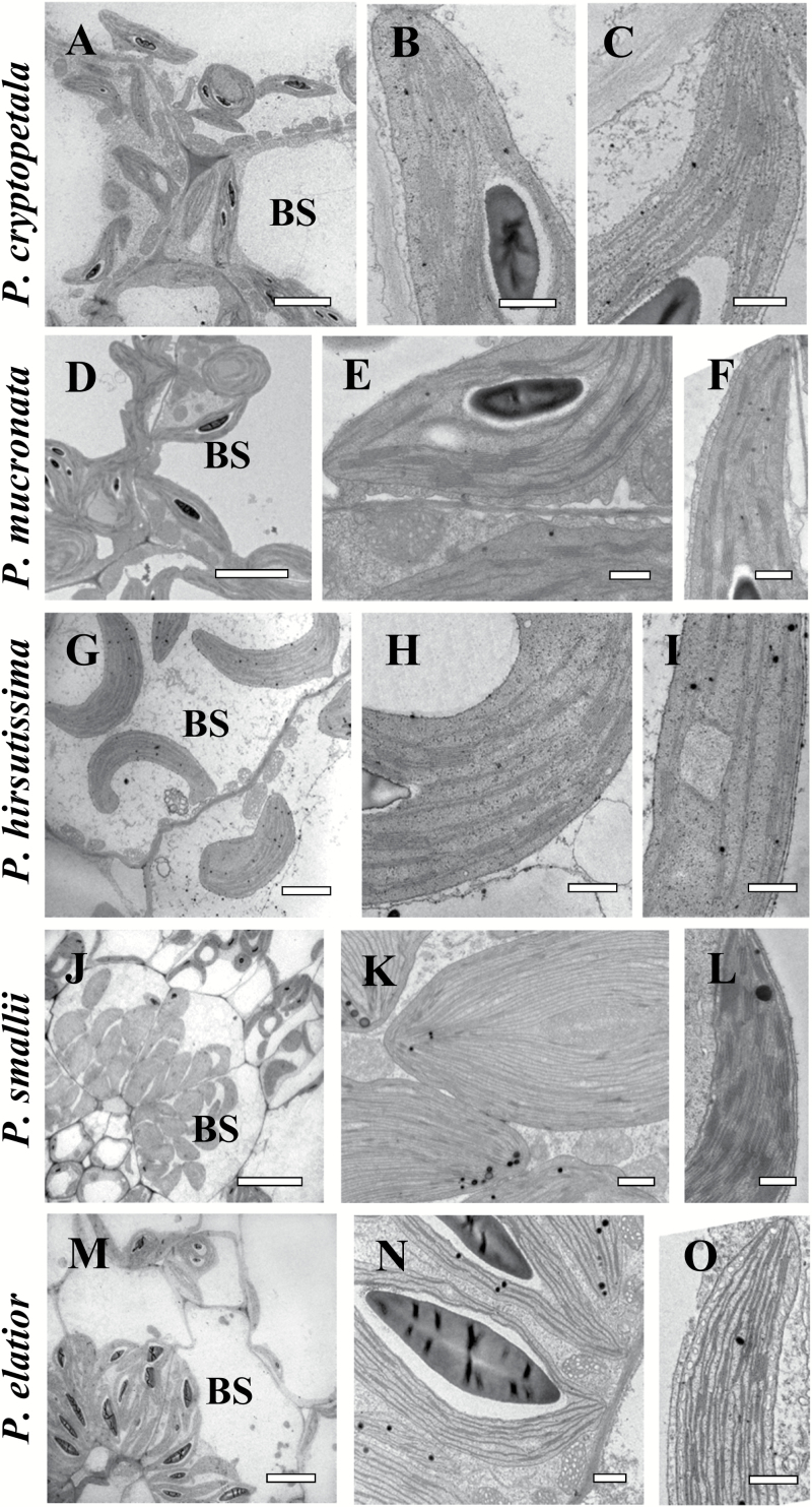
Electron microscopy of bundle sheath (BS) cells (A, D, G, J, M), and chloroplasts in BS (B, E, H, K, N) and mesophyll (M) (C, F, I, L, O) cells in representative Portulaca species. P. cryptopetala (A–C), P. mucronata (D–F), P. hirsutissima (G–I), P. smallii (J–L), and P. elatior (M–O). Panels (A, D, G, J, M) show the distribution of organelles in BS cells in centripetal position. In panels (B, E, H, N) the BS chloroplasts have well-developed grana, while BS chloroplasts in P. smallii (K) are grana-deficient. In contrast to the developed grana in M chloroplasts in (C, F, I and L), panel O shows grana-deficient M chloroplasts in P. elatior. Panel N illustrates occurrence of mitochondria around BS chloroplasts. BS, bundle sheath; M, mesophyll;. Scale bars: 5 µm for A, D, G, M; 20 µm for J; 1 µm for K; 0.5 µm for B, C, E, F, H, I, L, N, O.

