Fig. 6.
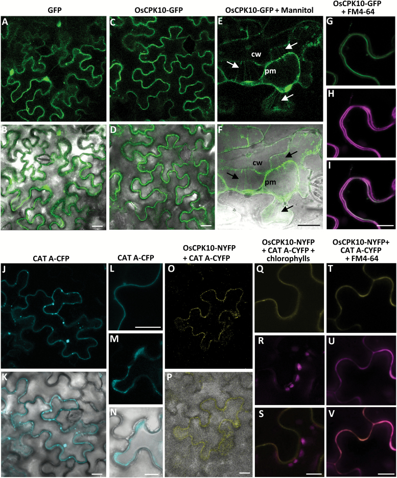
OsCPK10 is localized at the cell plasma membrane and interacts with CAT A. Confocal fluorescence microscopy of N. benthamiana epidermal cells transformed with GFP (A, B), OsCPK10-GFP (C–I), CAT-CFP (J–N), or OsCPK10-NYFP and CATA-CYFP (O–V) via Agrobacterium. Images were taken 48 h after agroinfiltration. (E, F) Plasmolysed cell after 15 min of treatment with mannitol. Arrows indicate the Hetchian strands attaching the plasma membrane (pm) to the cell wall (cw). (H, U) Transformed cell stained with the lipophilic dye FM4-64. (A, C, E, G–J, L, M, O, Q–V) Fluorescence images. (B, D, F, K, N, P) Fluorescence and bright field merged images. (I) Merged image of green (G) and magenta (H) fluorescence. (R) Chlorophyll autofluorescence (magenta signals). (S) Merged image of the yellow fluorescence corresponding to the reconstituted YFP and the chlorophyll autofluorescence (magenta). (V) Merged imaged of yellow (T) and magenta (U) fluorescence. Scale bars, 10 µm.

