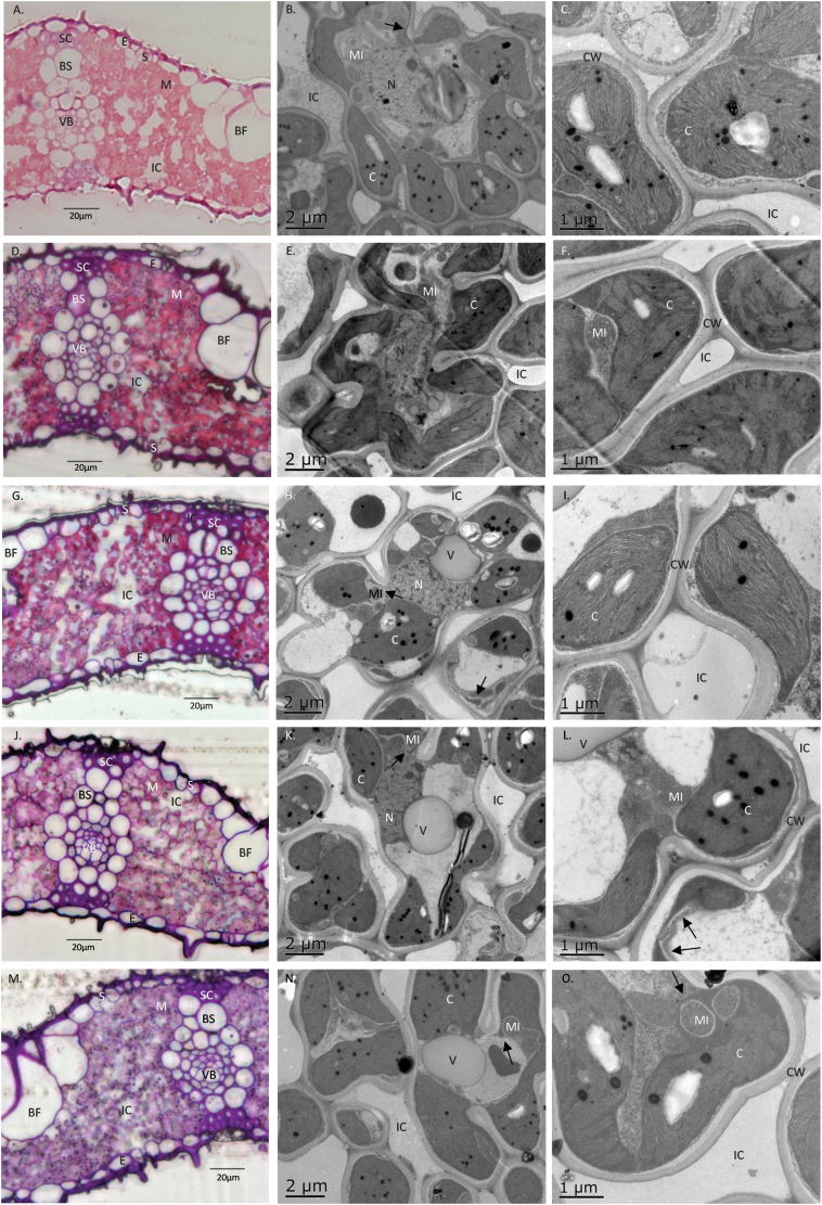
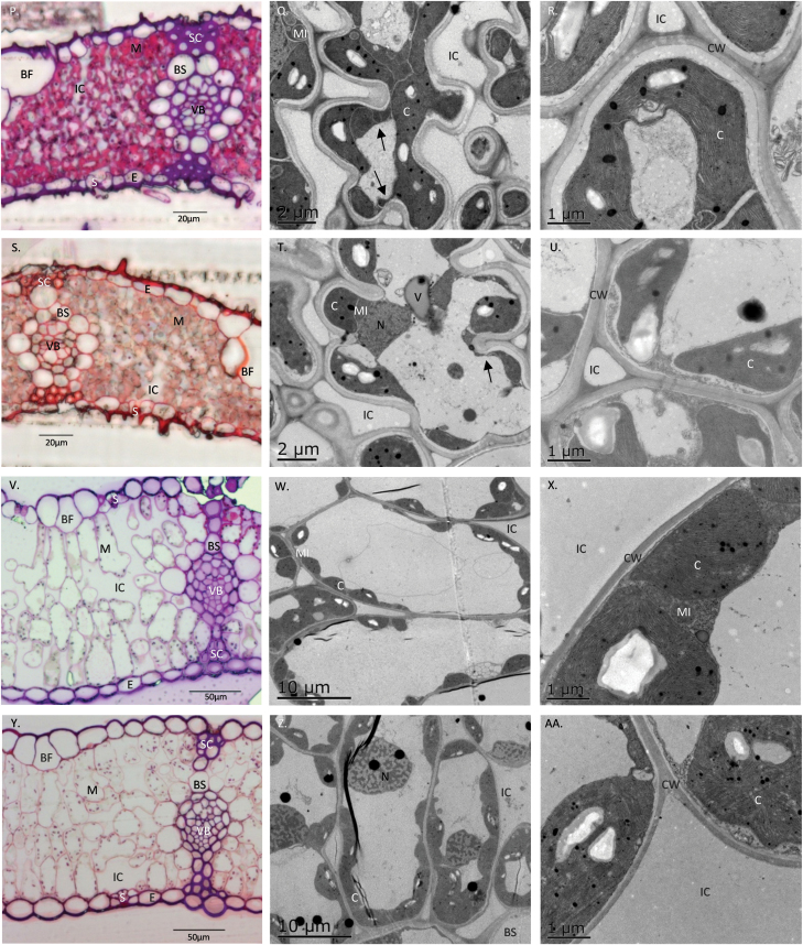
Fig. 6.
Light (left panels) and electron (middle and right panels) micrographs of leaf structure and anatomy for O. sativa cultivars (A–C, IR64; D–F, II; G–I, Apo; J–L, 148; M–O, UPL7; P–R, Sal), O. glaberrima (S–U, CG14), and T. aestivum cultivars (V–X, S82; Y–AA, We4). The left panels show the overall leaf transverse sections; the middle panels show mesophyll cell shape, chloroplast distribution, and lobe development; and the right panels show mesophyll cell walls. Arrows mark stromules. BF, bulliform cell; BS, outer bundle-sheath cell; C, chloroplast; CW, mesophyll cell wall; E, epidermis; IC, intercellular airspace; M, mesophyll cell; MI, mitochondria; N, nucleus; S, stoma; SC, sclerenchyma strand; VB, vascular bundle; V, vacuole.


