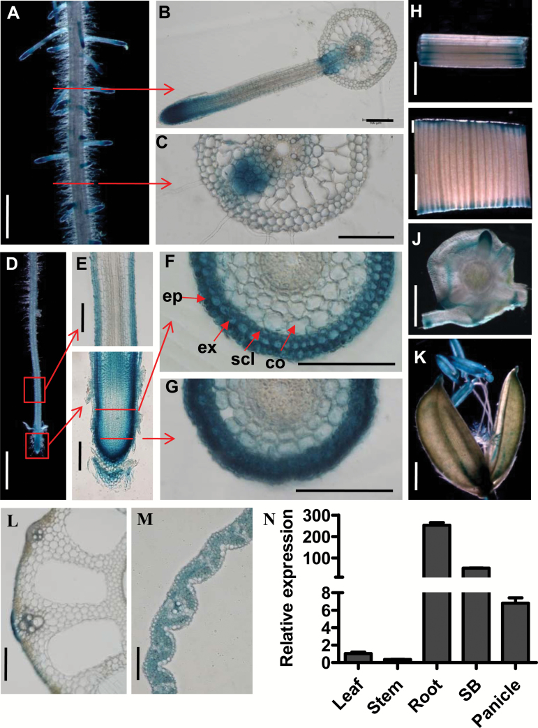
Fig 3.
Tissue-specific expression patterns of OsPIN2. (A–M) GUS staining of transgenic plants harboring the OsPIN2p:GUS reporter gene. (A) Mature root region with lateral roots, (B) cross-section of the region where a lateral root emerges, (C) cross-section of the region where a lateral root primordium is initiated, (D) primary root tip, (E) longitudinal section of primary root tip (lower panel) and the elongation zone (upper panel), (F) cross-section of the elongation zone, (G) cross-sections of a root cap, (H) stem, (I) leaf, (J) stem base, and (K) panicle. Abbreviations: ep, epidermis; ex, exodermis; scl, sclerenchyma; co, cortex. Scale bars indicate 1 mm in (A, D, H–K), 100 µm in (B, C, E–G, M), 200 µm in (L). (N) Quantitative RT-PCR analysis of OsPIN2 mRNA levels in the leaf, stem, root, stem base (SB), and panicle. The primers used are listed in Supplementary Table S1.

