Fig 4.
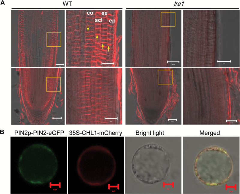
Tissue-specificity and subcellular localization of OsPIN2. (A) Tissue-specificity in the root tip. Immunostaining was performed with an anti-PIN2 antibody in root tips of the wild-type (WT) and lra1 (longitudinal sections). The boxed areas are magnified in the images to the right. Abbreviations: ep, epidermis; ex, exodermis; scl, sclerenchyma; co, cortex. Arrows indicate polar localization of OsPIN2. Scale bars =20 µm. (B) Subcellular localization of OsPIN2. The OsPIN2-GFP fusion protein driven by the OsPIN2 promoter and CHL1-mCherry (cell membrane marker) driven by the 35S promoter were transiently co-expressed in rice protoplasts. Fluorescent signals as observed by confocal microscopy are shown. Scale bars =5 µm.

