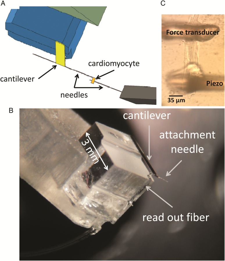
Figure 1.
The experimental approach. (A) Schematic view of the experimental set-up. (B) Microscope image of an intact, live cardiomyocyte glued in between the two anchoring needles; one of the two needles is anchored to the free hanging end of the cantilever, the other to a piezoelectric translator that allows one to modulate the length of the cell. (C) Microscope image of the force sensor.

