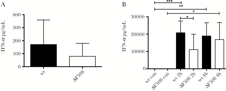
Figure 3.
ΔF508 mice show a delayed interferon-alpha (IFN-α) response after stimulation with poly(I:C). A, Wild-type (wt; n = 6) and ΔF508 (n = 8) mice were infected with Coxsackievirus B3 (104 plaque-forming units [PFU]/mouse) and serum was drawn on day 2 postinfection. B, Wt (n = 8–14 per time point) and ΔF508 (n = 6–8 per time point) mice were stimulated with poly(I:C) (100 μg/mouse, intraperitoneal) and serum was drawn at 2 hours and 4 hours after stimulation. IFN-α levels in serum from wt mice (black bars) and ΔF508 mice (white bars) from infected mice (A) or mice stimulated with Poly(I:C) (B) were measured using enzyme-linked immunosorbent assay. Data is presented as means ± SD *P < .05, **P < .01, and ***P < .001, two-way analysis of variance with Holm–Sidak’s correction.

