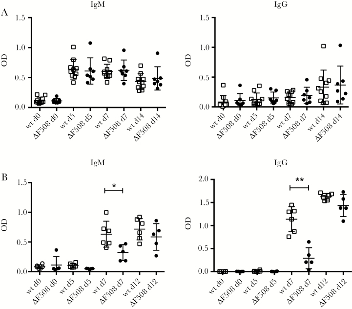
Figure 6.
ΔF508 mice have an intact T-cell–independent antibody response but show a delayed response to a T-cell–dependent antigen. A, Wild-type (wt; n = 10; black circles) and ΔF508 (n = 7; open squares) mice were injected with the T-cell–independent antigen 2,4,6-trinitrophenyl (TNP)-Ficoll (10 µg/mouse, intraperitoneal [IP]). B, Wt (n = 6; black circles) and ΔF508 (n = 5; open squares) mice were challenged with the T-cell–dependent antigen recombinant Semliki Forest virus expressing beta-galactosidase (rSFV-βGal; 1 × 10⁵ Infectious units [IU]/mouse, intravenous [IV]). Serum was drawn at the indicated time points (days 0, 5, 7, and 12 postinjection) and levels of nitrophenyl (NP)-specific antibodies in serum (A) and βGal (B) specific IgM and IgG were measured using enzyme-linked immunosorbent assay. Open squares and black circles represents wt and ΔF508 mice, respectively. Data are presented as mean ± standard deviation *P < .05, **P < .01. Mann−Whitney test. Abbreviation: OD, optical density.

