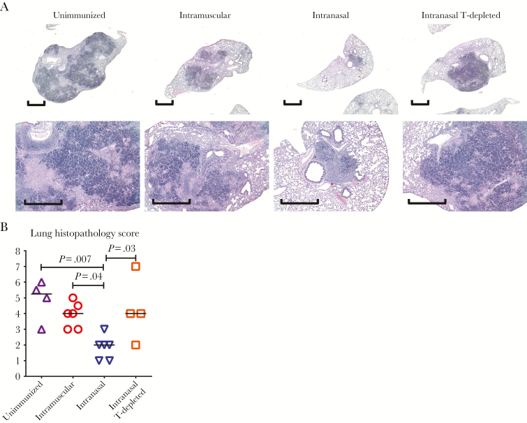
Figure 7.
Pulmonary tuberculosis histopathologic findings in immunized humanized mice (Hu-mice). Unimmunized animals, animals immunized parenterally (intramuscularly) or via the respiratory mucosa (intranasally), and animals immunized intranasally and depleted of human CD4+ and CD8+ T cells (T-depleted) were infected with Mycobacterium tuberculosis (1 × 10⁴ colony-forming units per animal) via the pulmonary route 4 weeks after viral-vectored immunization and euthanized 4 weeks after infection. A, Representative micrographs of lung sections stained with hematoxylin–eosin, comparing the extent of granulomatous lesions and necrosis. Scale bar represents 1 mm. B, Scatterplot comparing the scores of microscopic histopathologic changes in the lung. Horizontal lines in B represent the median values, and data in B are pooled from 2 independent experiments with the indicated number of animals per group.

