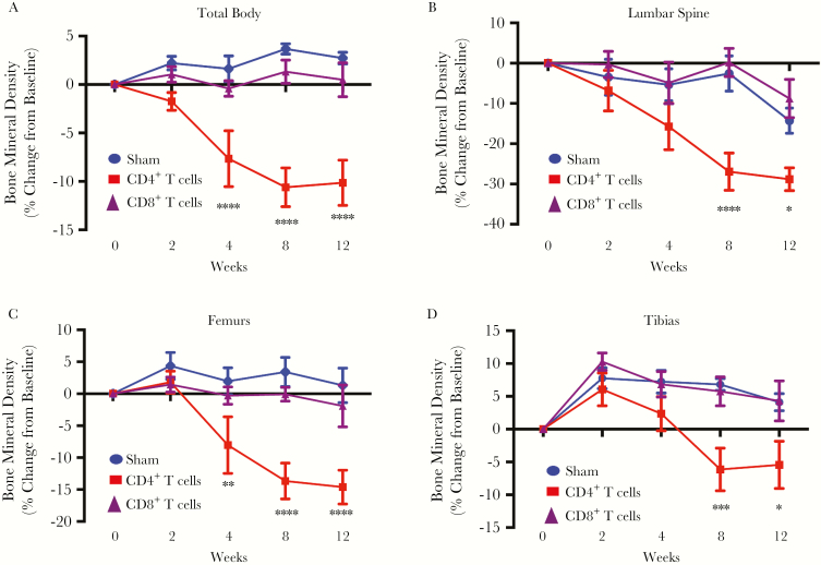
Figure 1.
Prospective bone mineral density in mice transplanted with CD4+ or CD8+ T cells. Bone mineral density (BMD) total body (A) and in the lumbar spine (B), femur (average of left and right femur for each mouse; C), and tibia (average of left and right tibia for each mouse; D) was quantified by dual-energy x-ray absorptiometry at baseline (0 weeks) and 2, 4, 8 and 12 weeks following CD4+ T-cell or CD8+ T-cell adoptive transfer. All data are expressed as means ± standard errors of the mean. Data are for 12 mice in the sham group and 6 mice each in the CD4+ and CD8+ T-cell–reconstituted groups. *P < .05, **P < .01, ***P < .001, ****P < .0001, compared with the sham group, by 2-way analysis of variance with the Tukey multiple comparisons post hoc test.

