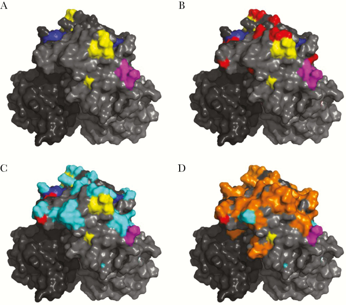
Figure 1.
GII.17 capsid P2 domain residue changes over time. A, Structural model of GII.17.2002 P domain dimer (shades of gray) with GII.4 surface-exposed blockade epitope A (yellow), D (blue), and E (magenta) highlighted. B–D, Residue changes between 1978 and 2002 (red; B), between 2002 (cluster I) and 2005 (cluster II; cyan; C), and between 2005 (cluster II) and 2015 (cluster IIIb; orange; D).

