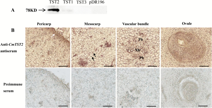
Fig. 6.
Immunolocalization of CmTST2 in melon. (A) Western blot of yeast-expressed CmTST2 to test the specificity of the anti-CmTST2 antiserum. A specific 78-kD band was detected only in cells expressing CmTST2 and was absent in cells expressing CmTST1, CmTST3, or the pDR196 empty vector. (B) Transverse sections of melon fruit at 0 d after anthesis. Xyl, xylem; Ph, phloem. The white arrow indicates the epicarp; black arrows indicate the mesocarp cells. Scale bars =100 μm. (This figure is available in colour at JXB online.)

