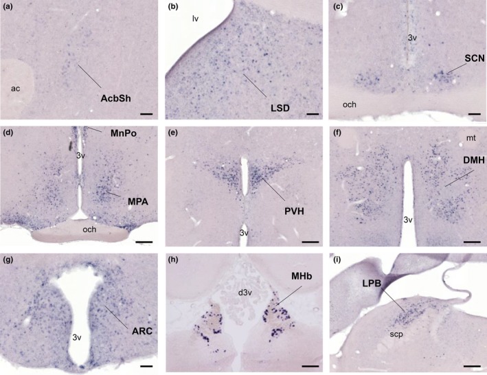
Figure 3.

Distribution of BRS‐3 mRNA in the rat CNS. (a–i) Coronal rat brain sections were stained with a rat BRS‐3 antisense cRNA probe. Results and abbreviations are summarized in Table 3. Bar: 500 μm (a–c, g), 250 μm (d–f, h, i). 3v, 3rd ventricle; ac, anterior commissure; d3v, dorsal 3rd ventricle; mt, mammillothalamic tract; lv, lateral ventricle; och, optic chiasm; scp, superior cerebellar peduncle
