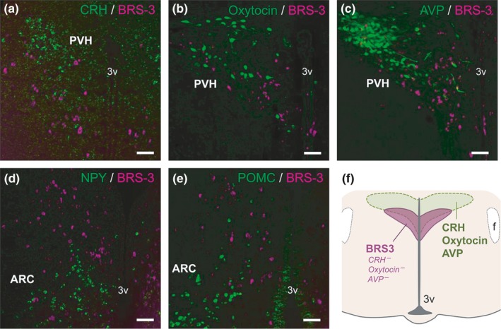
Figure 4.

Double staining of BRS‐3 mRNA with various feeding‐related neuropeptides in the rat paraventricular hypothalamic nucleus (PVH) and ARC. (a–c) Coronal rat brain sections of the PVH showing BRS‐3 mRNA (magenta) and (a) CRH mRNA signals, and (b) oxytocin‐ir signals and (c) AVP‐ir signals (green). (d, e) Coronal rat brain sections of the ARC (bregma −3.7 mm) showing BRS‐3 mRNA (magenta) and (d) NPY mRNA signals and (e) POMC mRNA signals (green). f, Schematic representation showing the localization of the BRS‐3‐expressing neuronal cluster in the PVH. Bar: 50 μm. 3v, 3rd ventricle; f, fornix
