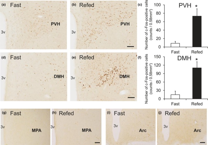
Figure 5.

Hypothalamic c‐Fos induction in the rats refed for 2 hr after 48 hr of food deprivation. Coronal brain sections from rats fasted for 48 hr (Fast) or refed for 2 hr (Refed) were stained with anti‐c‐Fos antibody. (a–f) c‐Fos induction in the paraventricular hypothalamic nucleus (PVH) (a–c) and dorsomedial hypothalamic nucleus (DMH) (d–f) following refeeding. a, b, d, and e show representative photographs indicating the c‐Fos‐ir signals in PVH (a and b, bregma −1.9 mm) and DMH (d and e, bregma −3.2 mm). c and f show the calculated density of c‐Fos‐ir cells in the 0.58‐mm2 area. Average number of the total c‐Fos‐ir cells in each animal was follows; 35 ± 18 (PVH, Fast), 293 ± 70 (PVH, Refed), 60 ± 50 (DMH, Fast), and 441 ± 87 (DMH, Refed). Results are presented as mean values ± standard deviation (n = 3). *p < .05 versus vehicle (Student's t test). (g–j) Photographs showing c‐Fos‐ir signals in the MPA (g and h, bregma −0.3 mm) and ARC (i and j, bregma −3.8 mm). Bar: 100 μm. 3v, 3rd ventricle; MPA, medial preoptic area
