Figure 6.

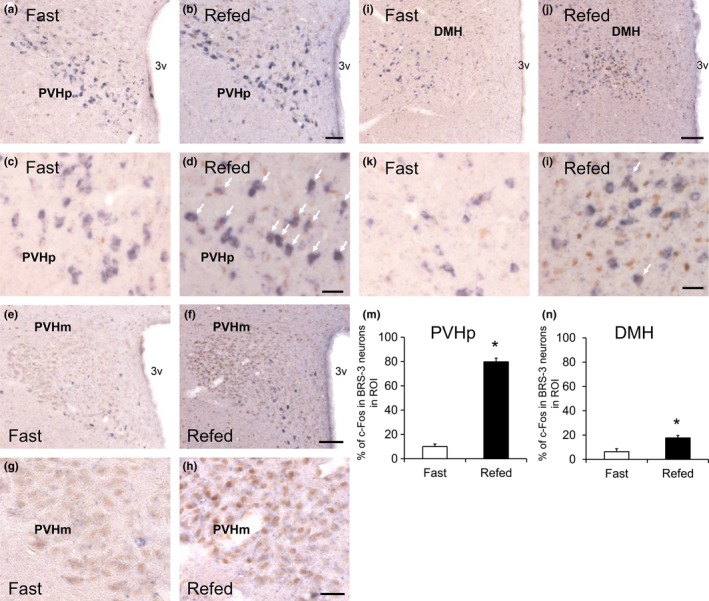
c‐Fos induction in the BRS‐3 mRNA‐positive neurons of rats refed for 2 after 48 h of food deprivation. (a–l) Coronal brain sections from rats fasted for 48 hr (Fast) or refed for 2 hr (Refed) were stained with rat BRS‐3 antisense cRNA probe and anti‐c‐Fos antibody. BRS‐3 mRNA‐positive signals (purple) and c‐Fos‐ir signals (brown) were detected in the paraventricular hypothalamic nucleus (PVH)p (a–d, bregma −1.9 mm), PVHm (e–h, bregma −1.7 mm), and dorsomedial hypothalamic nucleus (DMH) (i–l, bregma −3.2 mm). c and d, g and h, and k and l show high magnification of PVHp (a and b), PVHm (e and f), and DMH (i and j), respectively. The white arrows show double‐positive cells for BRS‐3 mRNA and c‐Fos‐ir signals. Bar: 50 μm (a and b), 25 μm (c, d, g, h, k, and l), and 100 μm (e, f, i, and j). 3v, 3rd ventricle. (m–n) The percentage of c‐Fos‐ir signals in the BRS‐3 mRNA‐positive neurons (percentage of c‐Fos in the BRS‐3 neurons in ROI) within the PVHp (m) and DMH (n) was calculated from the images; 114.3 ± 19.1 (PVHp) and 100.3 ± 10.3 (DMH) of BRS‐3 neurons in each animal were examined. Results are presented as mean values ± standard deviation (n = 3). *p < .05 versus vehicle (Student's t test)
