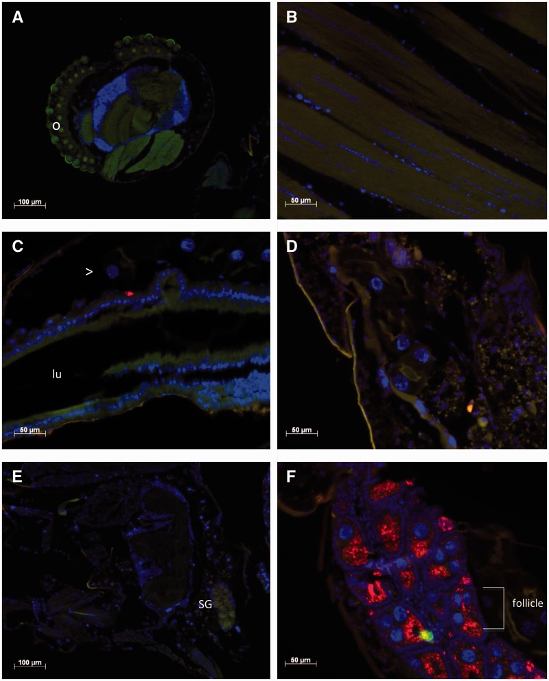
Fig. 2.
Localization of Wolbachia in Ae. notoscriptus. Fluorescence in situ hybridization (FISH) of paraffin sections showing the localization of Wolbachia (red) in different tissues of 7-d-old female Ae. notoscriptus. Wolbachia are labeled using two 16 S rRNA rhodamine-labeled probes. DNA is stained with DAPI (blue) and a green GFP filter is used to provide contrast. ( A ) Head. “o” indicates the ommatidia that form the compound eye of the mosquito. ( B ) Thoracic muscle. ( C ) Midgut. The red spot is an artifact and not Wolbachia. “Lu” indicates the midgut lumen and “>“ the malpighian tubule running parallel to the midgut. ( D ) Malpighian tubule. ( E ) Salivary gland, labeled “SG”. ( F ) Ovary showing the presence of Wolbachia (red).

