. 2017 Sep 25;8(12):8025–8029. doi: 10.1039/c7sc03772d

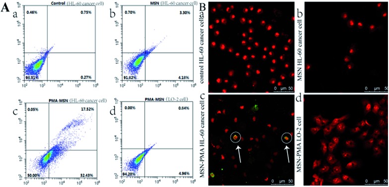
Fig. 4. (A) Flow cytometry study of HL-60 cells (0.5 mL, 1 × 10⁶ cells per mL) after incubation with (a) PBS buffer, (b) MSNs and (c) PMA@MSN probe (1 mg mL–1) for 12 h and stained with the Annex V-FITC/PI apoptosis kit. (d) Flow cytometry study of LO-2 cells (0.5 mL, 1 × 10⁶ cells per mL) after incubation with PMA@MSN probe (1 mg mL–1) for 12 h and stained with Annex V-FITC/PI. (B) Merged confocal microscopy images of the TUNEL-FITC/PI staining of HL-60 cancer cells after treatment with (a) PBS buffer, (b) MSNs and (c) PMA@MSN probe for 12 h, respectively. (d) Merged confocal microscopy images of LO-2 cells incubated with PMA@MSN probe for 12 h and stained with TUNEL-FITC/PI.

Fig. 4