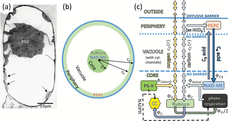
Fig. 1.
Modelling a Bienertia mesophyll cell. (a) Micrograph of a mature Bienertia mesophyll cell, taken from Voznesenskaya et al. (2002) with permission. The arrows point to the peripheral chloroplasts. (b) Model of a Bienertia cell, showing the three compartments, and marking the location of various enzymes. The compartment’s radii, ri, rv, and re are varied in the model, so the picture should not be taken to scale. (c) Abstract schematic of reactions and flows in different spatial regions considered in the model. Yellow arrows represent the oxygen current, and light blue is the CO2 current (two-headed arrows represent diffusion). Other arrows represent the C3 and C4 acid currents (grey and dark blue), and the photorespiratory and Calvin–Benson cycle carbon currents (striped green and blue). The thin dashed line is the NADPH current originating from the Hill reaction in the core’s chloroplasts; it couples the photorespiratory and the Calvin–Benson cycle activity to the oxygen production. The boundary between the periphery and the outside is the only barrier to gas diffusion in the model.

