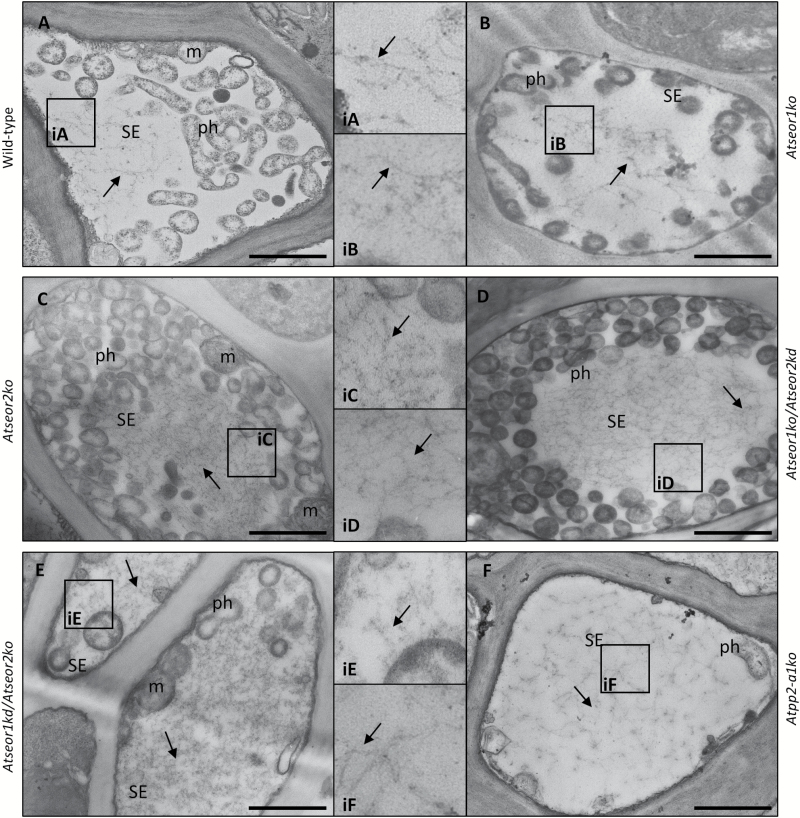
Fig. 12.
TEM micrographs of sieve elements in different infected A. thaliana lines. Different A. thaliana lines were indicated as follows: (A) wild-type, (B) Atseor1ko, (C) Atseor2ko, (D) Atseor1ko/Atseor2kd, (E) Atseor1kd/Atseor2ko, and (F) Atpp2-a1ko. In infected tissue, wild-type (A) and Atpp2-a1ko (F) SEs showed a massive presence of SE protein filaments (arrows, insets). Also in A. thaliana lines lacking one or both AtSEOR genes (B–E), infected SEs showed filaments with morphology and organization similar to those observed in wild-type plants (arrows, insets). For each condition, five non-serial sections from 10 different plants were analysed. CC, companion cell; m, mitochondrion; ph, phytoplasma; SE, sieve element. Scale bars: 1 µm.

