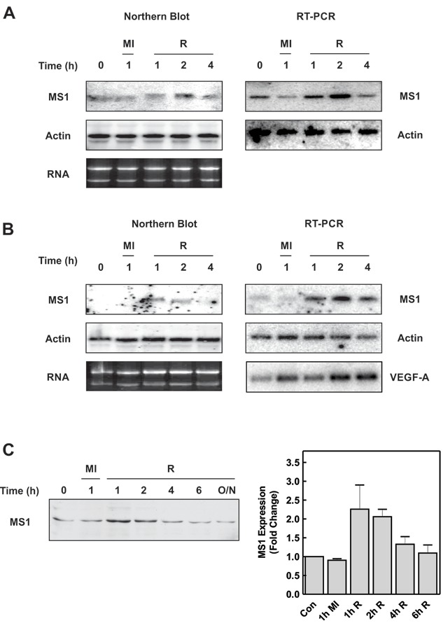
Figure 1.

MS1 expression is increased during metabolic stress in cardiac myocytes. Neonatal cardiac myocytes (A) or H9c2 cells (B) were subjected to metabolic inhibition (MI) or allowed to recover (R) following 1h of metabolic inhibition for the times indicated. Northern blots for MS1 and β-Actin mRNA are presented on the left-hand side with MS1, VEGF-A and β-Actin mRNA determined by RT-PCR on the right. Ethidium bromide-stained gels showing 28S and 18S RNA levels are also shown. (C) Cell extracts from H9c2 cells subjected to metabolic inhibition (MI) and recovery (R) were blotted with antibodies to MS1. A representative blot and a graph indicating quantification of the data are shown (mean ± SEM [n = 3]).
