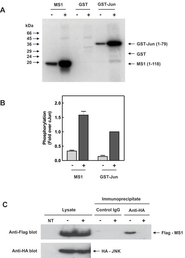
Figure 4.

MS1 is a JNK target. (A) HEK cells, transfected with pcDNA3-HA-JNK and expressing HA-tagged JNK, were incubated with (+) or without (–) 0.5 M sorbitol for 30 minutes to activate the JNK pathway. HA-JNK was immunoprecipitated from the cell lysates and incubated with [γ-32P] ATP along with a purified N-terminal fragment of MS1 (1–118), GST or GST-Jun (1–79) in an in vitro immune-complex kinase assay. 32P-incorporation into substrates was determined by SDS-PAGE and PhosphorImager analysis. (B) Quantification was performed using ImageQuant software and the relative phosphorylation of MS1 compared to GST-Jun is shown (mean ± SEM [n = 3]). (C) HEK cells were co-transfected with pcDNA3-HA-JNK and pFlag-CMV2-MS1 to express both HA-tagged JNK and Flag-tagged MS1. The cells were treated with 0.5 M sorbitol for 30 min (+) or left untreated (–) and cell lysates immunoprecipitated with anti-HA antibody or control IgG. Cell lysates and immunoprecipitates were separated by SDS-PAGE and blotted with anti-Flag and anti-HA antibodies. Non-transfected cell lysates were run as a negative control (NT).
