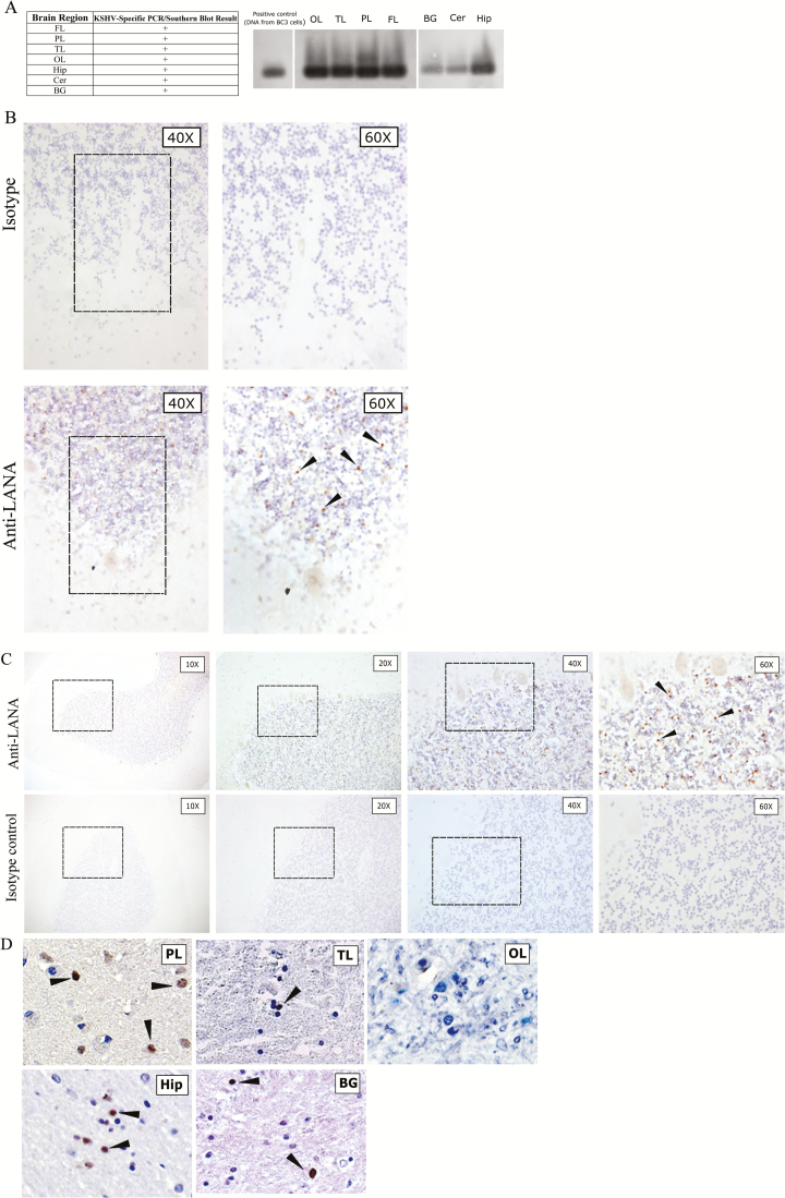
Figure 1.
A, Southern blot hybridization–confirmed polymerase chain reaction (PCR) results for a human immunodeficiency virus (HIV)–positive, Kaposi’s sarcoma (KS)–associated herpesvirus (KSHV)–seropositive, but KS-asymptomatic individual (subject 116), showing that most central nervous system (CNS) tissues harbored KSHV genomic DNA. Southern blot with probe against KSHV ORF26 is shown. Demographic and clinical characteristics of subject 116 are available in Table 1. B, Findings of immunohistochemical (IHC) analysis of CNS tissue specimens from subject 116, using near-adjacent cerebellum (Cer) sections for isotype and anti–latency-associated nuclear antigen (LANA) staining. The different magnifications are of the same cerebellar folium. LANA-positive cells appeared as brown punctate staining, and representative cells are designated by arrowheads. C, Progressive magnifications of anti-LANA IHC staining of Cer sections from subject 116 (top) in comparison to isotype control IHC staining of a near-adjacent section (bottom). The architecture of the folia is evident at lower magnification, and the Purkinje cells bordering the granular layer are resolved at higher magnifications. Boxes denote the field magnified in the subsequent panel. LANA-positive cells appeared as brown punctate staining, and representative cells are designated by arrowheads. D, IHC staining of addition brain regions from subject 116 reveals KSHV LANA in the parietal lobe (PL), temporal lobe (TL), hippocampus (Hip), and basal ganglia (BG) but not in the occipital lobe (OL). LANA-positive cells appeared as brown punctate staining, and representative cells are designated by arrowheads. Isotype control staining of the same tissue specimens was negative (not shown). Control staining with anti-LANA in a KSHV-seronegative subject is shown in Supplementary Figure 1. ART, antiretroviral therapy; +, positive.

